Due to its unpredictable nature, cancer growth and progress in an individual is difficult to determine. As a cancer grows and progresses in a body, it also alters the effectiveness of the treatment and therefore chances of survival. In simple terms, the earlier you detect cancer, by and large the greater your chances of surviving the disease.
It is possible to detect certain cancers before they begin to show symptoms in an individual. The process of checking for cancer in people who have no symptoms is called screening. Screening is mostly effective as it helps in early and more effective treatment of some cancers. As symptoms appear the cancer could become harder to treat. For cancer… “The best protection is early detection.”
DETECT CANCER EARLY
There are some cancers for which screening is possible. Screening is the ability to detect a cancer in an individual before it has begun to show symptoms. The female cancers (breast and gynaecological) for which screening is possible are:
A clinical breast examination is a physical examination of the breast done by a health professional to check for breast changes or breast problems.
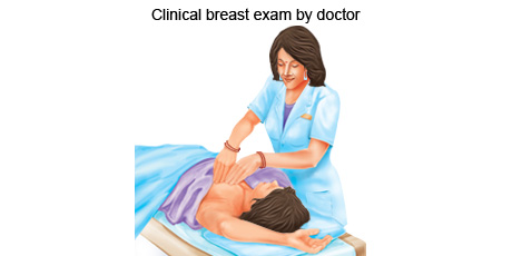
A clinical breast examination is done by a doctor or health professional where the doctor carefully feels the breasts and under the arms for lumps or anything else that seems unusual. The procedure normally does not cause any discomfort.
The health professional then examines each breast, underarm, and collarbone area for changes in breast size, skin changes, or signs of injury or infection, such as bruising or redness. The individual may be asked to lift their arms over their head, put their hands on their hips, or lean forward and press their hands together to tighten the muscle beneath each breast during this part of the examination. They may also lie flat on the table and put their arm behind their head while the health professional checks the breast tissue.
The health professional will feel each breast for any unusual or painful areas or for a lump. The health professional will gently press on the breast tissue from about 1 inch (2.5 cm) below the breast up to the collarbone. She will also examine the armpit and your neck for swollen glands. The health professional will likely press gently on your nipple to check for any discharge.
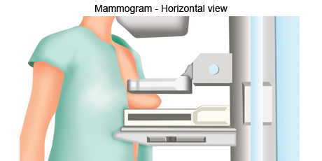
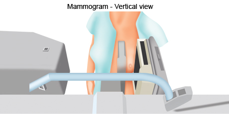
A mammogram is a low-dose X-ray exam of the breasts that allows one to have a closer look for changes in breast tissue that cannot be felt during a breast exam.
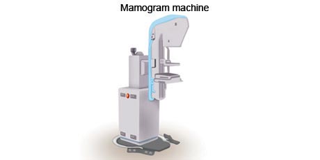
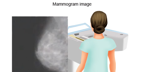
Mammography is a technique that takes low dose x-ray images of breasts. It is done using a mammography machine. Mammography has the ability to find tumours that may be too small to feel during a physical exam. The ability of a mammogram to find breast cancer depends on the size of the tumour, the density of the breast tissue, and the skill of the radiologist. Mammograms are less likely to find breast tumours in women younger than 40 years than in older women. This may be because younger women have denser breast tissue that appears white on a mammogram. Given a tumour also appears white on a mammogram, it makes it harder to differentiate between the two.
During a mammogram each breast is pressed between two metal plates. This is done to flatten the breast as much as possible so a clear image can be taken of the breast with a low dose of x-ray. Having the breast pressed in this manner may be uncomfortable but this only lasts a few minutes. The x-ray image is then taken from above. This process is repeated for the second breast. It is important to be still while the image is being taken. The plates of the mammography machine will then be moved around to take an image from a different angle. The process of mammography could last up to 30mins.
A breast self examination is a physical examination of the breast done by an individual to check for changes or abnormalities in the breast. This is not a screening test but a useful self assessment tool.
A breast self examination is a systematic step-by-step approach to examine the look and feel of ones breasts to detect any signs and symptoms of breast problems like a lump, skin irritation, dimpling, nipple retraction (turning inward) or discharge other than breast milk. The exam is done monthly between the 7th and 10th day of a woman's menstrual cycle when the female hormones are mostly in balance. The aim of the breast self exam is to detect breast cancer early.
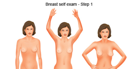
The breast self examination is a 5 step process.
- Breasts that are their usual size, shape, and colour
- Breasts that are evenly shaped without visible distortion or swelling
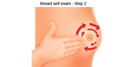
Step 2 Stand and press your three middle fingers to your breast, working around the breast in a circular direction using your right hand to feel your left breast and then your left hand to feel your right breast. Use a firm, smooth touch with the finger pads of your hand, keeping the fingers flat and together. Use a circular motion, about the size of a 1 Rupee coin. Follow a pattern to be sure that you cover the whole breast. You can begin at the outer edge of the breast, moving in smaller circles until you reach the nipple.
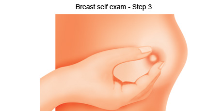
Step 3 Squeeze your nipples to check for discharge. This could be a watery, milky, or yellow fluid or blood. In case of a bloody discharge, see your doctor immediately.
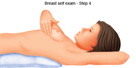
Step 4 Next, feel your breasts while lying down. Place a pillow or folded towel under your left shoulder. This helps your breast tissue spread evenly across your chest wall. Now repeat Steps 2 and 3.
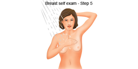
Step 5 Finally, feel your breasts while you are in the shower. It is easier to feel your breasts when the skin is wet and slippery. Cover your entire breast, using the same hand movements described in Step 2.
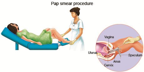
A pap smear is also called a cervical smear and checks for changes in the cells of the cervix (the organ connecting the uterus and vagina) that are taken by taking a sweep of the opening of the cervix.

Regular screening for women 21 years and older with the Pap test decreases their chance of dying from cervical cancer. In women younger than 21 years, screening with the Pap test may show changes in the cells of the cervix that are not cancer but lead to further testing and possibly treatment. The Pap test is only recommended for sexually active women.
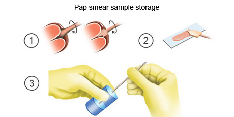
It is recommended that a Pap test be done in the middle of the menstrual cycle. A Pap test is a procedure to collect cells from the surface of the cervix and vagina. A piece of cotton, a brush, or a small wooden stick is used to gently scrape cells from the cervix and vagina. The cells are viewed under a microscope to find out if they are abnormal. This procedure is also called a Pap smear. A new method of collecting and viewing cells has been developed, in which the cells are placed into a liquid before being placed on a slide. This allows for transportation of the sample to distant location for analysis.
Not all types of cancer have screening tests and some tests are only for people with specific genetic risks. Luckily, screening is possible for the two most common female cancers – breast cancer and cervical cancer. Screening is also possible for oral cancer which is done as a visual inspection by a doctor.
It is important to remember that when your doctor suggests a screening test, it does not always mean he or she thinks you have cancer. Screening tests are done when you have no cancer symptoms.
